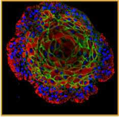
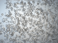
Reproductive system organoid models in health and disease

Reproductive System

Principal Investigator
- prof. dr. Hugo Vankelecom
- Department of Development and Regeneration
- Cluster of Stem Cell and Developmental Biology, Unit of Stem Cell Research
- Laboratory of Tissue Plasticity in Health and Disease
- Campus Gasthuisberg - O&N4 – 5th floor (room 05.239)
- KU Leuven
- Hugo.vankelecom@kuleuven.be
- https://www.kuleuven.be/samenwerking/scil/hugovankelecom
Short CV
Hugo Vankelecom is PhD in Pharmaceutical Sciences and teaches Pharmacology and Stem Cell Biology at KU Leuven. His group is highly proficient in developing, characterizing and applying organoid models, a booming field but still quite unique at KU Leuven. The team developed and applied organoid models from endometrium and endometrial diseases to delve into the mechanisms underlying the (patho-)biology of this essential reproductive organ and embryo implantation; from the pituitary, the master gland in the neuroendocrine-reproductive axis, to decipher its (patho)biology, its still mysterious stem cells and regeneration; and from tooth to understand mouse/human tooth stem cells, development, disease, regeneration and repair.
3D tissue models
- Organoid and assembloid models (human and mouse) from:
- endometrium to study its biology and deficient function in gynecological and reproductive diseases (endometriosis, polycystic ovary syndrome (PCOS), cystic fibrosis, …)
- endometrium to study embryo-endometrium interaction in a human implantation model \
- endometrial cancer
- ovarian cancer and ovary
- reproductive organs to enable drug discovery/personalized medicine


Methods – expertise
Established organoid lines are amplified, cryopreserved for biobanking and subjected to downstream characterization and application processes
- Assays: healthy and diseased tissue organoids used to study molecular and cellular (patho-)physiology (e.g. hormonal responsiveness, single-cell omics, …), embryo implantation, and for drug discovery and screening.
- Imaging: histochemical and immunofluorescence staining analysis of paraffin sections, and high-end imaging of full-3D immunofluorescently stained organoids (confocal, light-sheet, live recording/Incucyte ,…).
- live culture imaging of organoid growth and behavior (Incucyte).
Application
- Unraveling the biology of healthy (human) endometrium
- Disease modeling of endometriosis, PCOS and cystic fibrosis
- Modeling and unravelling embryo-endometrium interaction
- Drug discovery and screening
- Personalized medicine
Some key publications
- Boretto, M. et al. Development of organoids from mouse and human endometrium showing endometrial epithelium physiology and long-term expandability. Development 144, 1775–1786 (2017). 10.1242/dev.148478
- Boretto, M. et al. Patient-derived organoids from endometrial disease capture clinical heterogeneity and are amenable to drug screening. Nature Cell Biology 21, 1041–1051 (2019). 10.1038/s41556-019-0360-z
- Maenhoudt N, Defraye C, Boretto M, Jan Z, Heremans R, Boeckx B, Hermans F, Arijs I, Cox B, Van Nieuwenhuysen E, Vergote I, Van Rompuy AS, Lambrechts D, Timmerman D, Vankelecom H. Developing organoids from ovarian cancer as experimental and preclinical models. Stem Cell Reports, 14:717-729 (2020). 10.1016/j.stemcr.2020.03.004.
- Kagawa, H. et al. Human blastoids model blastocyst development and implantation. Nature 601, 600-605 (2022). 10.1038/s41586-021-04267-8
Collaboration opportunities
- Mouse and human healthy and diseased endometrium-derived organoid lines
- Reproductive biology and diseases using organoid models
- Embryo-endometrium interaction using a human implantation model
- Reproductive tract cancers (endometrium, ovary)
Contact person
- prof. dr. Hugo Vankelecom

